1. Trachomatous inflammation follicular (TF)
Overview
The examination of the eye for trachoma begins with observation of the cornea and the eyelids for corneal opacity and in-turned eyelashes. The upper eyelid must then be everted in order to observe the superior tarsal conjunctiva.
This is the conjunctiva that covers the stiffer part of the eyelid and occupies the central region. The conjunctiva at the lateral and medial margins and over the rounded edge of the tarsal plate should not be examined. Only the central region is examined because some of the signs that are graded are normal findings outside this region.
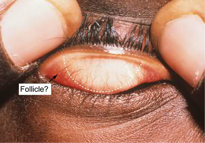
Figure 5. Grading area.
The simplified WHO grading system looks for the presence or absence of five signs: each sign is assessed independently of the other signs. An eye may not have any signs, it may have only one sign, it may have all five or a combination in between. Follicles must only be considered if they lie within the grading area white dashed line. Follicles can occur outside this region as part of the normal anatomy. Considering these follicles when grading may lead to an incorrect diagnosis of TF in a normal patient.
Trachomatous inflammation follicular (TF)
Definition
The presence of five or more follicles 0.5 mm diameter in the upper tarsal conjunctiva.
TF is one of the signs of active inflammatory disease or infectious trachoma. The other is trachomatous inflammation intense (TI). Each sign is assessed independently of the other signs and should only be considered if clearly present.
Always use the rule: If in doubt leave it out.
For the TF sign to be considered the following conditions must be met. Five or more follicles of at least 0.5 mm diameter must be present in the grading area. Follicles are lymphoid aggregates that represent the body’s immune response against C. trachomatis. They appear as round well demarcated white areas that are paler than the surrounding conjunctiva. Small scars and degenerative deposits in the conjunctiva can be confused with follicles and care must be taken to avoid this. Small scars will generally not have a round border, being irregular in shape with sharp corners. Degenerative deposits can be yellow or white masses with clear cut edges. Cysts, appearing as clear bubbles should also be distinguished from follicles.
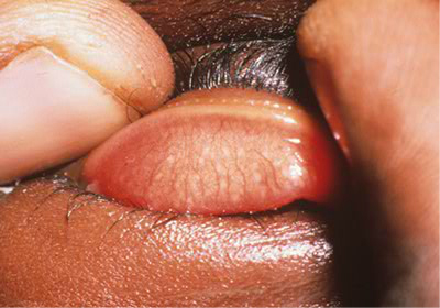
Figure 6. Trachomatous inflammation follicular (TF).
A quick examination of the grading area should be made, and 5 follicles should be identified. If there is doubt as to whether a follicle is present or not, it should be left out. It is important that an eye with only 4 follicles is not graded as TF.
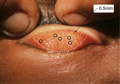
Figure 7: Diagram demonstrating the size of a 0.5 mm follicle in proportion to the rest of the eye.